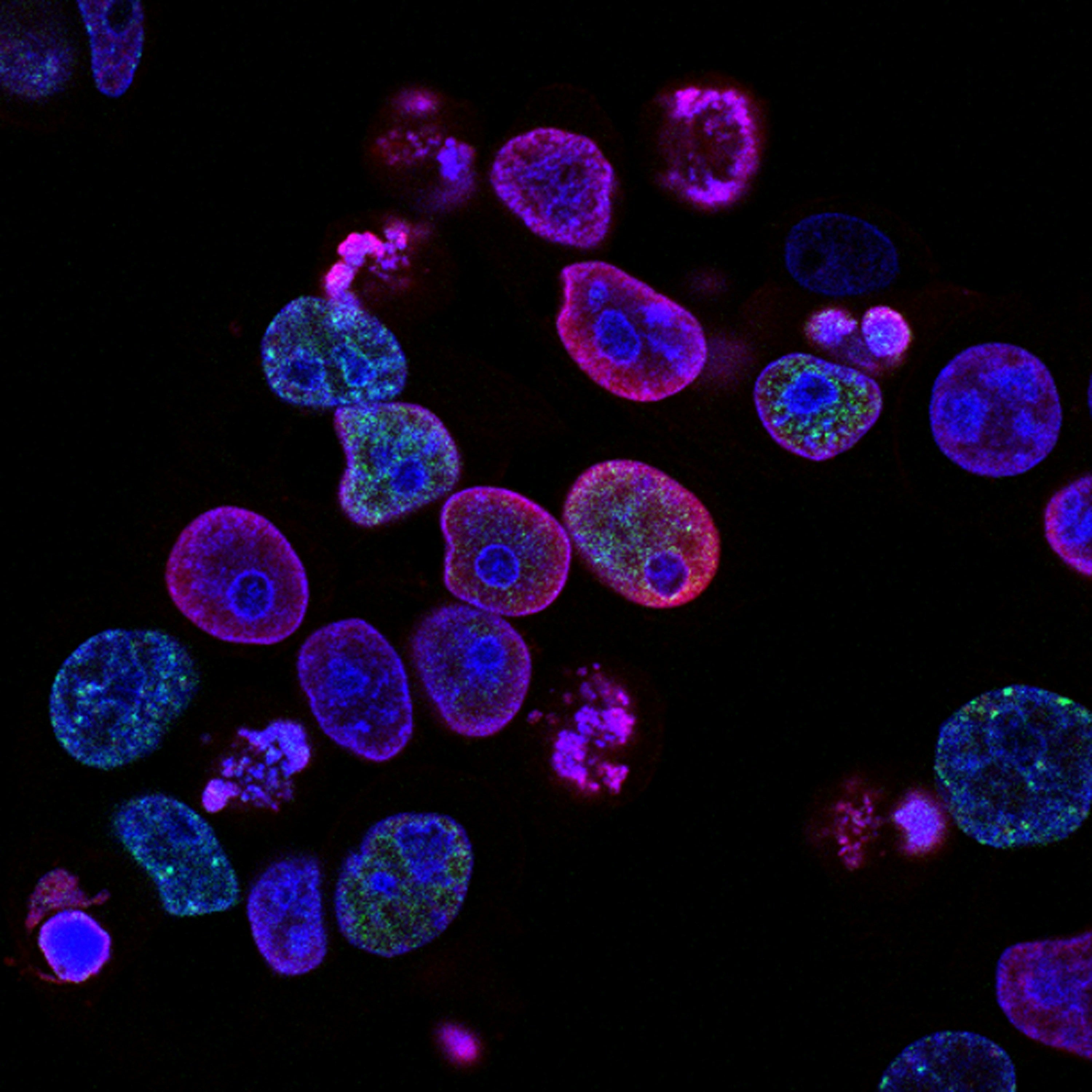
ChromaLIVE non-toxic dye goes beyond cell painting to reveal cellular phenotypes in high-content screening
Cell images hold high-density information about cell states and their responses to drugs. However, existing in-vitro assays capture only limited information and offer a simplified view of cell response. Fortunately, with ChromaLIVE, scientists can now reveal much more of the precious information contained in cells.
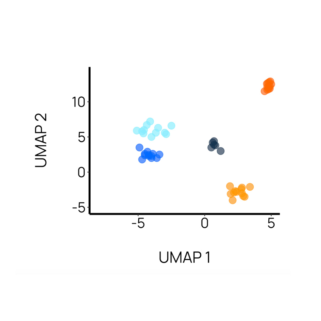
ChromaLIVE, the leading technology for image-based profiling, generates phenotypic signatures that allow differentiating between drugs or genetic perturbations. It enables scientists to gain better understanding of biological events or drug mechanism at higher throughput than - and with similar biological relevance as - alternative profiling methods like RNA sequencing.

ChromaLIVE Features
Data-rich dye for new cell insights
Non-Toxic
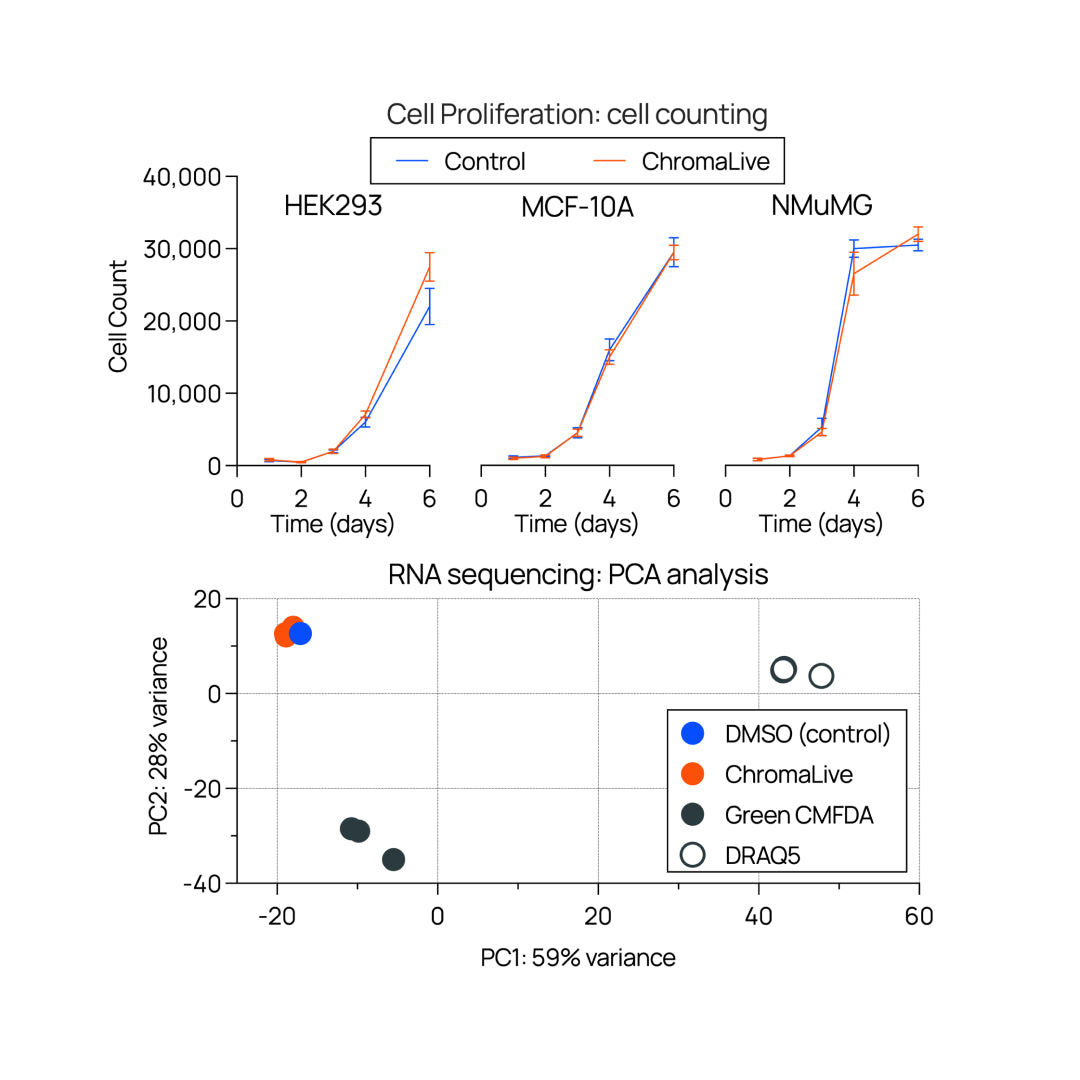
Unaffected gene expression patterns and stable in multi-week live cell cultures.
Mix-and-read
One-step, no-wash dye which does not fluoresce in media. Remains in culture media throughout the assay.
Data-rich
ChromaLIVE alone provides high-density biological information, and therefore helps differentiate subtle phenotypic signatures

Applications
New insight you didn’t know you could get
Check Out Our Resources


NucleoLIVE: non-toxic nuclear dye protocol for live-cell imaging

MortaLIVE: non-toxic dye protocol for live-cell imaging

MortaLIVE™: A non-toxic dye for live-cell imaging and real-time cytotoxicity assays
Frequently Asked Questions
-
RNA sequencing data and week-long cell cultures support this claim. Figure 2 of this SLAS Discovery article presents supporting RNA sequencing and proteomics data showing unperturbed expression patterns – in contrast with alternative live cell dyes – for cells cultured in presence of ChromaLive.
In addition, ChromaLive has been successfully tested in sensitive cell systems without affecting cell health for weeks on end. For instance, it has been used in a 3-week culture of iPSC-derived neurons, and a 6-week culture of patient-derived prostate cancer organoids.
-
RNA sequencing data and week-long cell cultures support this claim. Figure 2 of this SLAS Discovery article presents supporting RNA sequencing and proteomics data showing unperturbed expression patterns – in contrast with alternative live cell dyes – for cells cultured in presence of ChromaLive.
In addition, ChromaLive has been successfully tested in sensitive cell systems without affecting cell health for weeks on end. For instance, it has been used in a 3-week culture of iPSC-derived neurons, and a 6-week culture of patient-derived prostate cancer organoids. -
ChromaLive is one product which is a multi-chromatic small molecule dye that is excited at two different wavelengths: 488nm, with a long-stokes shift emission in the red (making it compatible with your own GFP fluorophore) and 561nm which has standard yellow emission.
See our document “How does ChromaLive work” in our resources for more details such as how it stains multiple cell compartments at the same time: https://www.saguarobio.com/resources/
-
Fluorescence signal from ChromaLive can both be retained or removed after fixation, depending on your downstream assay needs. Signal retention requires our ChromaLive Fix additive and is performed with 4% PFA. By fixing and permabilizing cells (without ChromaLive Fix additive) signal can be entirely removed.
-
ChromaLive is multichromatic and is therefore excited at two wavelengths: 488nm and 561nm. The 488 channel has a long-stokes shift emission in the red (making it compatible with your own GFP fluorophore), while the 561 channel has standard yellow emission. Most high-content imaging instruments are compatible with ChromaLive. Refer to our resources on our website to find a list of compatible instruments, along with technical notes detailing filter selection specific to the Yokogawa, Molecular Devices and Revvity systems. Incucyte systems are also compatible with ChromaLive!
-
ChromaLive comes in tubes of 10uL, and is used at a dilution of 1:1,000 in culture medium. The biological inertness of ChromaLive and its fluorescence only upon incorporation into cells allow its addition at the cell seeding stage, which is highly recommended. Addition of ChromaLive at a later stage is possible, but it is recommended to test staining kinetics as optimal staining can take up to 12 hours depending on cell type.
-
Reach out for a quote! Volume pricing is available, and academic discounts too.

